
Fig. 2.
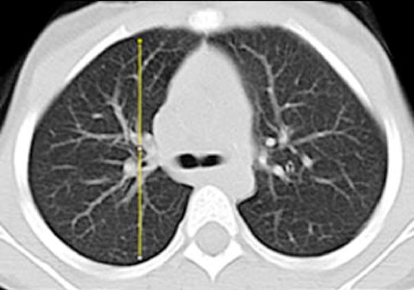
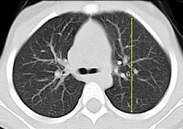
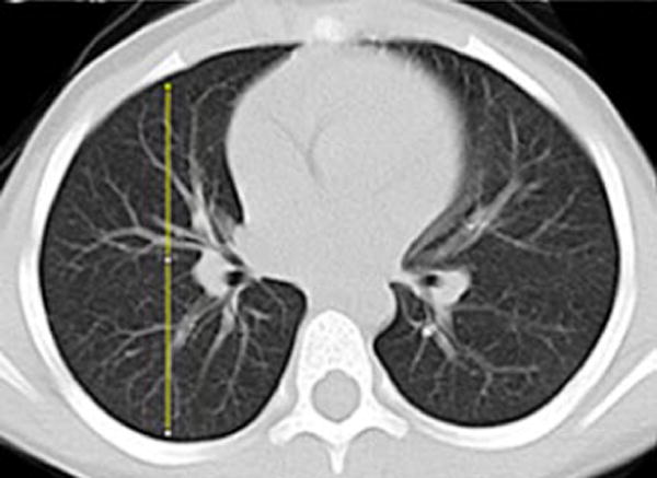
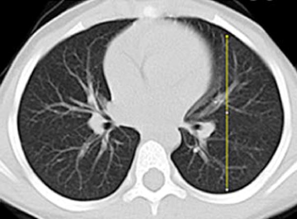
Image analysis of lung attenuation. Non-contrast axial CT images through the thorax in a 46-month-old boy demonstrate the method used to measure gravitational dependence of lung attenuation with ImageJ. Four linear plot profiles were obtained in each child, located in each upper lung at the level of the carina (a, b), and in each lower lung at the approximate level of the basilar segmental bronchi origins (c, d)