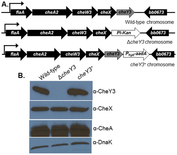
Figure 1. Construction and complementation of the ΔcheY3.
A. WT B. burgdorferi genome arrangement of flaA operon containing cheY3 (labeled as WT chromosome). The Pl-Kan (aph1) cassette was inserted after deleting the cheY3 gene by allelic exchange (ΔcheY3 chromosome). The mutant was complemented in cis by genomic reconstitution by inserting a WT copy of the cheY3 gene flanked by the PflgB-aadA cassette (cheY3+ chromosome). Arrows indicate the direction of transcription. DNAs/Plasmids are not drawn to scale. B. Immunoblot analysis of B. burgdorferi cells probed with the indicated antibodies. The CheY3 protein expression was inhibited in the mutant, but restored in the complemented cheY3+ cells as confirmed by using cell lysates from the indicated clones probed with anti-CheY3. CheY3 protein is approximately 14 kDa. The cheA2 and cheX genes are located in the same operon as the targeted cheY3, however, the expression of those gene products were not altered in the mutant or the complemented cells (see anti-CheA and anti-CheX blots). DnaK was used as a loading control.

