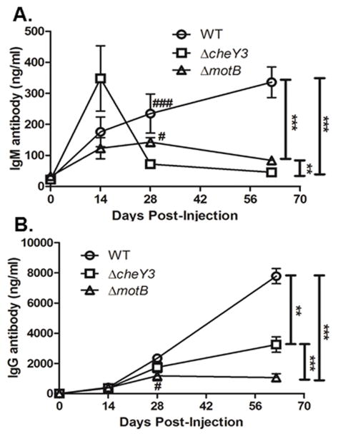
Figure 7. Infection with ΔcheY3 is sufficient to elicit B. burgdorferi-specific antibodies.
The mice used in the dissemination study (Figure 6) were sacrificed at the indicated time post-injection and sera were collected. B. burgdorferi-specific antibodies were quantified by ELISA analyses using bacterial sonicates as the capture antigen. A. Detection of IgM antibody levels in serum from WT-, ΔcheY3- and ΔmotB-infected mice. ΔmotB spirochetes was used as a control as these bacteria are reported to be non-motile, fail to disseminate from the injection site, and are cleared from the host within 48-72h after injection. **p < 0.01, ***p < 0.0001 as shown in the graph. # p < 0.05 and ### p < 0.0001 as compared to the ΔcheY3. Results were analyzed using the ANOVA test followed by the Tukey-Kramer Multiple Comparisons test. B. Detection of IgG antibody levels in serum from WT-, ΔcheY3- and ΔmotB-infected mice. **p < 0.01, ***p < 0.0001 as shown in the graph. # p < 0.05 as compared to WT. Statistical analyses were performed using ANOVA test followed by Tukey-Kramer Multiple Comparisons test.

