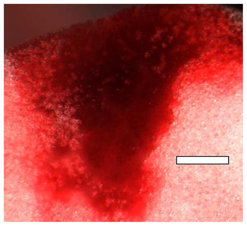
Fig. 5.

A PCH area induced by scanning at an on screen MI=0.9 at 7.6 MHz (image from the study reported in Miller et al. (2014)). The alveolar gas has greatly diminished in the PCH volume, but residual bodies of gas remain as bubbles of about 100 μm diameter or less. Scale Bar: 1 mm.
