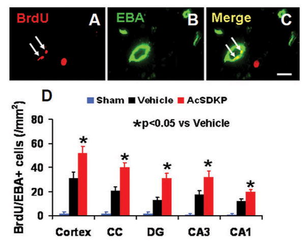
Fig. 4.

The effect of AcSDKP on the number of newly generated vessels in rats 35 days after TBI (8 rats per group). Double staining for BrdU (A, white arrows) and EBA (B, green signal) was performed to identify newly formed mature vessels (C, white arrows) in the brain at Day 35 after TBI in the lesion boundary zone and DG area. Scale bar = 20 μm (A–C). Data in the bar graphs (D) represent the mean ± SD.
