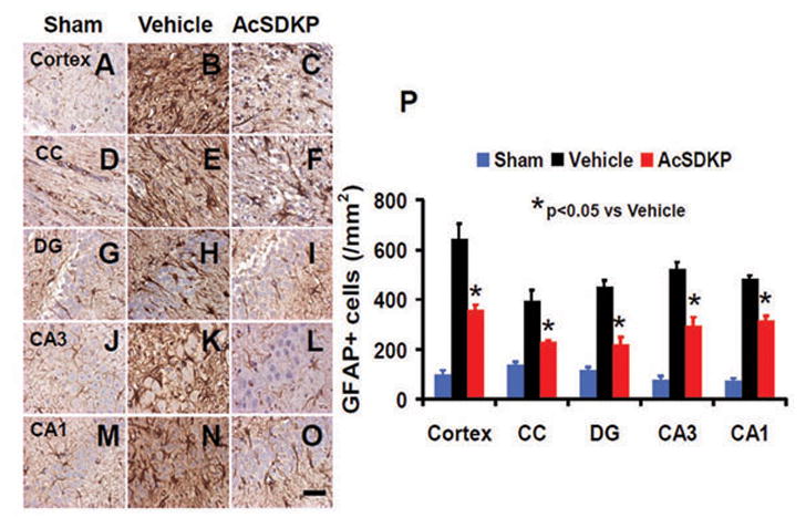
Fig. 8.

The effect of AcSDKP on astrocyte activation. GFAP staining was performed to detect activation of astrocytes 35 days after TBI. Some weak expression of GFAP was observed in brain regions of sham animals (A, D, G, J, and M). AcSDKP treatment (C, F, I, L, and O) significantly decreased GFAP-positive cells in various brain regions 35 days after TBI compared with the saline group where prominent astrogliosis exists (B, E, H, K, and N). CC = corpus callosum; DG = dentate gyrus. The data on GFAP-positive cells are shown in the bar graph (P). Scale bar = 20 μm. Data in graph represent mean ± SD. There were 8 rats per group.
