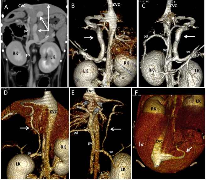
Fig. 5.
Splenophrenic PSS in dog 4 (A, B, C) and dog 10 (D, E, F) of CPSS group. (A) Dorsal multiplanar reformatted CT image of the abdomen at level of kidneys. A Splenophrenic PSS is evident (arrows) between splenic vein (sv) and post-hepatic segment of caudal vena cava (CVC). No varices nor abdominal effusion are evident. Three-dimensional volume-rendered CT angiography of portal system and caudal vena cava in dogs 4 and 10. (B, D – Dorsal views; C,E - ventral views; F - dorso-cranial view at level of hepatic surface.). On three-dimensional volume-rendered CT angiography of portal system and caudal vena cava of both dogs the splenophrenic PSS (arrows) had same anatomical pattern seen in dogs with PH of Fig. 1 and 2. These PSS were assumed to be congenital because appear as single porto-caval connections not associated with ascites or varices, and no structural causes of portal flow obstruction were evident on CT images.

