Fig. 1.

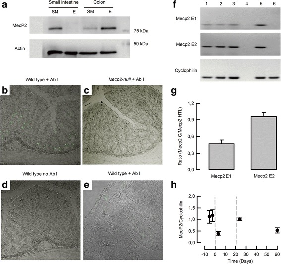
Protein and mRNA expression of Mecp2 in mouse intestine. a Representative western blot of Mecp2 from samples obtained from smooth muscle (SM) or epithelium (E) of the small intestine or colon of wild-type mice (n = 5). Mecp2 detection by immunofluorescence in b colon of wild-type, c colon of Mecp2-null mice, d colon of wild-type with omitted primary antibody, and e small intestine of wild-type mouse (n = 3 per group). f RT-PCR detection of Mecp2 variants E1 and E2 in colon epithelium. Lanes 1–3 colon samples, lane 4 RT-negative control, lane 5 hypothalamus sample from a wild-type mouse, and lane 6 hypothalamus sample from a Mecp2-null mouse. g Relative expression of Mecp2 variants on colon epithelium measured from the blots showed in f (n = 3). h qPCR from colon of embryonic 15.5 and 18.5 d.p.c. and 3, 24, and 60 days postpartum (n > 5 for each group)
