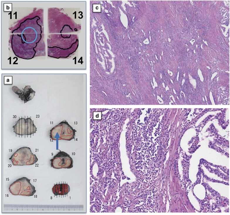
Figure 1.
Surgical specimens from case 7 in dose level-3 (DL-3) treated by one track injection with 1x10¹² vp of Ad-REIC. (a) Gross appearance of radical prostatectomy (RP) specimen sliced by a standard method. Cancer distribution areas are enclosed by the red line. A blue arrow indicates the Ad-REIC-targeted area. (b) Gross appearance of hematoxylin and eosin (H&E)-stained histopathological sections. Cancer distribution areas are enclosed by the black line. A blue circle indicates the targeted area. (c) A photomicrograph of the targeted area from section no. 12, demonstrating tumor degeneration with tumor-infiltrating lymphocytes (TILs) (H&E). (d) A photomicrograph of cancer distribution area from section no. 14, demonstrating a large intact tumor area (H&E).

