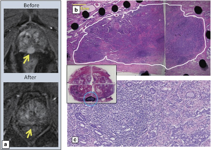
Figure 3.
Magnetic resonance imaging (MRI) and histopathological findings of case 13 in dose level-4 (DL-4) treated by three track injections with 3x10¹² vp of Ad-REIC. (a) Contrast enhancement MRIs. A strong enhancement area indicated by an arrow before Ad-REIC injections disappeared after injections. (b and c) Low and high magnification photomicrographs. Whole targeted cancer area is replaced by degenerated cancer cells with remarkable tumor-infiltrating lymphocytes (TILs). The inset shows gross appearance of hematoxylin and eosin (H&E)-stained histopathological sections.

