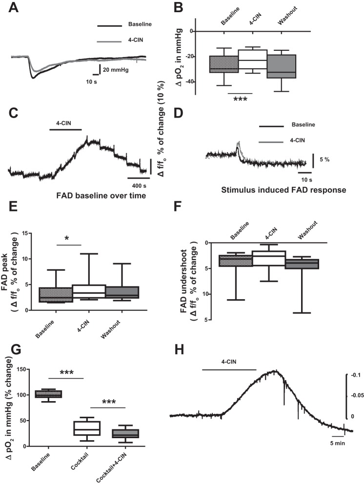
Fig. 5.
Effects of 4-CIN on partial oxygen tension (Po2) and FAD autofluorescence recorded in CA3 stratum pyramidale. A: sample traces of changes in stimulus-induced tissue oxygen tension before and during 4-CIN application. Note: reduction of change in stimulus-induced tissue oxygen tension. B: statistical analysis of effects of 4-CIN on ΔPo2. Note: ΔPo2 decreased during 4-CIN application and the effect was reversed during wash out. C: sample traces of stimulation-induced biphasic FAD/FADH2 response during 4-CIN perfusion. Note: the increase in FAD autofluorescence baseline during 4-CIN application. D: sample recordings of FAD autofluorescence are depicted before and during application of 4-CIN. E: statistical analysis of effects of 4-CIN on FAD fluorescence changes. The FAD peak significantly increased (F) while the undershoot was slightly reduced during 4-CIN application. G: statistical analysis of effects of 4-CIN on ΔPo2 associated with presynaptic action potentials. Presynaptic components of the stimulus-induced oxygen consumption was isolated by pharmacological blockade of postsynaptic glutamate and GABA receptors and presynaptic transmitters release. ΔPo2 decreased during co-application of the cocktail and 4-CIN. H: extracellular pH recording sample trace. Measurement of extracellular H+ ions was done using pH sensitive electrode; hence one observes an upward deflection with acidosis. Note: 4-CIN induced a decrease in extracellular pH. *P < 0.05, ***P < 0.001.

