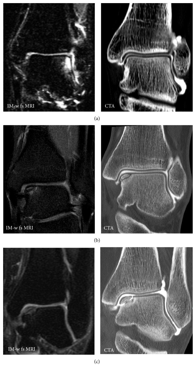
Figure 4.
Different presentations of osteochondral defects. (a) Patient with suspicion of an osteochondral defect on MRI but no defect on CTA. (b) Patient with an osteochondral defect without loosening (no subsequent surgery). (c) Patient with similar findings on MRI as in (b); however on CTA contrast enhanced fluid surrounds the osteochondral fragment indicating instability and the patient had to undergo subsequent surgery.

