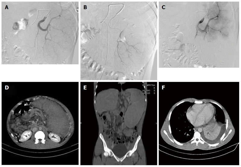
Figure 1.

Radiographic images of patient No. 10. A: Selective splenic arteriography showing the multiple splenic vessels supplying the enlarged spleen; B: Super selective catheterization of the inferior branches supplying the lower pole of the spleen; C: Post embolization images of the splenic artery, with no enhancement observed in the lower pole due to the embolized inferior lobe branch; D: Post procedural 1-mo follow-up with axial CT images, with no enhancement observed in the lower embolized portion of the spleen; E: Pre-procedural coronal reformatted CT images showing the enlarged spleen; F: Post-procedural axial CT image showing left pleural effusion accompanying lower lobe atelectasis.
