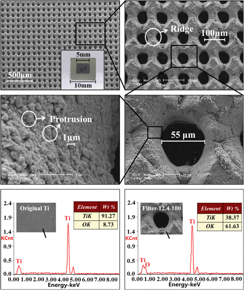
Figure 1. Surface morphology and chemical composition of filter-12.4-100.
(a–d) SEM images of ablated titanium foil fabricated with a laser fluence of 12.4 J/cm2 and a microhole spacing of 100 μm (filter-12.4-100). The inset in (a) is the photograph of filter-12.4-100. (a) Large-area view of filter-12.4-100. (b) Further magnified image of filter-12.4-100. (c) Enlarged view of a single microhole on filter-12.4-100. (d) Higher magnification image of the microhole wall. (e) EDXS result of the original Ti surface. (f) EDXS result of the ablated area on filter-12.4-100.

