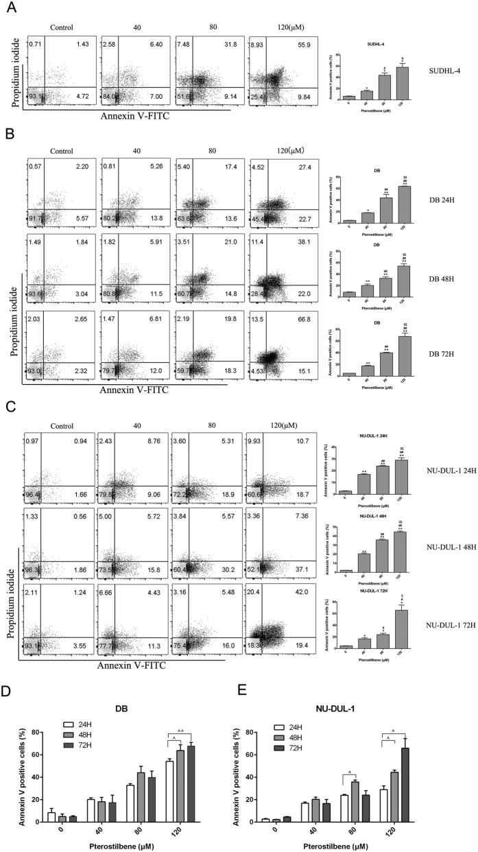
Figure 3. Pterostilbene increases apoptosis in DLBCL cell lines.
(A) SUDHL-4 cells were treated with various concentrations of pterostilbene (40, 80 and 120 μM) for 48 h, then cell viability was analyzed using flow cytometry. (B) DB and (C) NU-DUL-1 cells were treated with pterostilbene (40, 80 and 120 μM) at different time points (24, 48 and 72 h). Apoptosis was detected using a FITC-Annexin V/PI staining kit and examined by flow cytometry. The apoptotic index was expressed as the number of apoptotic cells/total number of cells counted × 100%. Columns represent the average percentage of Annexin V positive cells from three independent experiments, which are shown as the means ± SEM. *p < 0.05, compared with control groups; **p < 0.01, compared with control groups; #p < 0.05, compared with the pterostilbene 40 μM group; ##p < 0.01, compared with the pterostilbene 40 μM group; $$p < 0.01, compared with the pterostilbene 80 μM group. (D) DB and (E) NU-DUL-1 cells were exposed to pterostilbene at different time points (24, 48 and 72 h) after which the percentage of apoptotic cells was determined by Annexin V analysis. Data show means ± SEM. P values were calculated using one-way ANOVA. ^p < 0.05, compared with the pterostilbene 24 h group; ^^p < 0.05, compared with the pterostilbene 24 h group.

