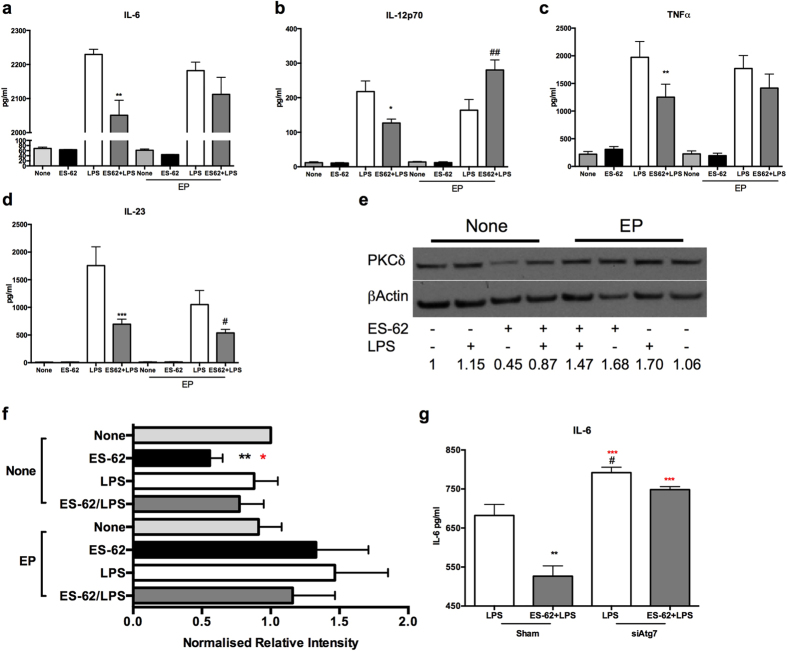
Figure 7. Blockade of autophagic flux prevents ES-62-mediated inhibition of LPS-stimulated IL-6, TNFα and IL-12p70 but not IL-23 production by DCs.
DCs were incubated in medium (None) ± ES-62 for 18 h and then with medium ± LPS for a further 18 h in the presence and absence of E64d plus pepstatin A (EP) and the levels of IL-6 (a), IL-12p70 (b), TNFα (c), and IL-23 (d) released, shown. Data in (a) are from a single experiment (means ± SD, n = 3) whereas in b-d, data are the means ± SEM of means collated from three independent experiments, where *p < 0.05, **p < 0.01 and ***p < 0.001 relative to the LPS group and #p < 0.05 and ##p < 0.01 relative to the LPS-EP group. DCs were incubated in medium ± ES-62 for 18 h and then medium ± LPS for a further 18 h in the presence and absence of EP and PKC-δ expression determined by Western Blot analysis (e) with quantitation of PKC-δ expression in this experiment relative to None/None control shown on the figure. Analysis was of data collated from 5 independent experiments for the None (no inhibitor) cohorts and 3 independent experiments for the EP cohorts (f) with data presented as means ± SEM where **p < 0.01 for ES-62 versus None and red*p < 0.05 for ES-62 versus LPS. In g, WT bmDCs were treated with control or ATG7-specific siRNA and then cultured in medium ± ES-62 prior to stimulation with LPS as indicated and then the levels of IL-6 measured by ELISA. Data are presented as means ± SD, n = 3 from a single experiment where **p < 0.01 is relative to the LPS-Sham, red***p < 0.001 relative to ES-62+LPS-Sham and, #p < 0.05 relative LPS-sham.

