FIG 4.
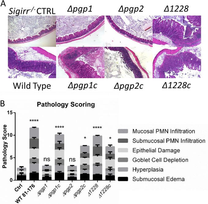
Tissue pathology in C. jejuni-infected Sigirr−/− mice. (A) Hematoxylin-and-eosin-stained histological sections from infected mouse ceca. Tissue samples were collected 7 days postinfection, formalin fixed, and paraffin embedded. Images were taken under ×100 magnification and were representative of tissues from at least seven mice. The uninfected control and Δpgp1 and Δpgp2 mutant-infected mice showed few signs of inflammation, whereas the ceca of mice infected with wild-type, Δ1228, and Δpgp1c, Δpgp2c, and Δ1228c complemented strains showed significant edema, crypt hyperplasia, and immune cell infiltration. (B) Pathological scoring was done by two blinded observers using hematoxylin-and-eosin-stained, formalin-fixed, cecal tissue sections. Scoring was done on a scale from 0 to 24, as described elsewhere (18). Significant pathology relative to uninfected controls was observed in mice infected with the wild type, the Δ1228 strain, and all complemented strains. No significant pathology was observed in mice infected with the Δpgp1 and Δpgp2 mutant strains. Statistical significance was determined using a two-way analysis of variance and a Bonferroni posttest. ns, not statistically significant; *, P > 0.05; ****, P > 0.0001.

