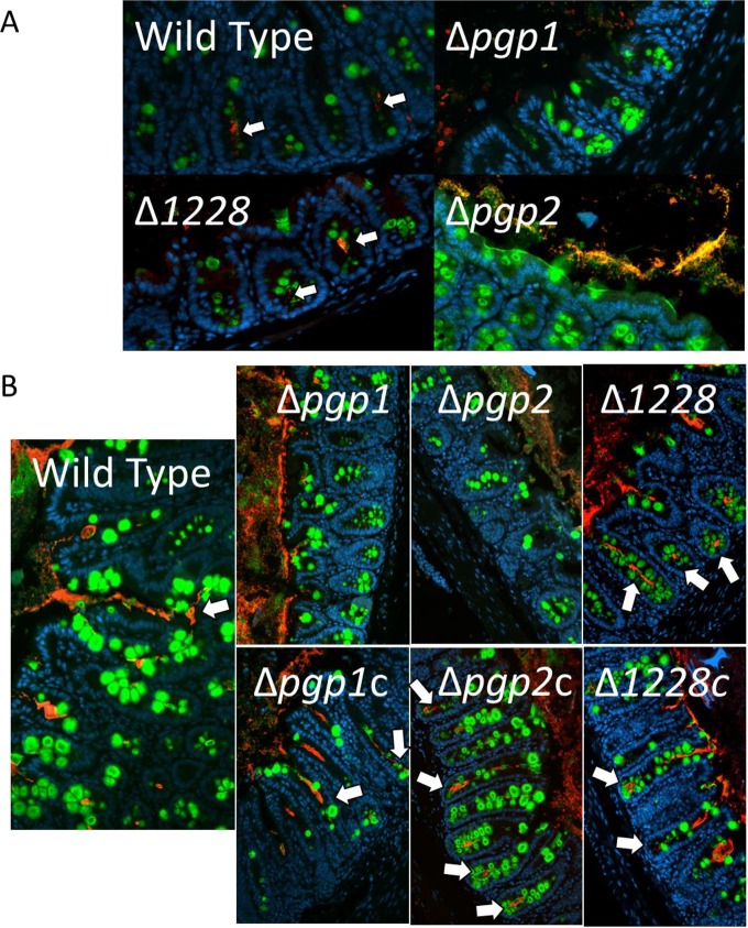
FIG 5.
Immunofluorescent staining of C. jejuni cecal colonization in infected Sigirr−/− mice. Formalin-fixed, paraffin-embedded tissue sections of ceca obtained from Sigirr−/− mice infected with wild-type, mutant, or complemented strains of C. jejuni at 3 days postinfection (A) or 7 days postinfection (B), at ×200 magnification. Cell nuclei are stained with DAPI (blue), the goblet cells and secreted mucus are stained with antibodies specific to Muc2 (green), and C. jejuni is stained with C. jejuni-specific antibodies (red). The wild type, the Δ1228 mutant, and all complemented strains are visible in the lumen, mucus layer, and crypts (white arrows). The Δpgp1 and Δpgp2 mutants are absent from the crypts.

