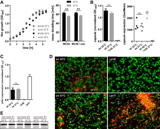
FIG 1.
Phenotypic characterization of N. meningitidis grown at 32°C or 37°C. (A) Growth curves of capsulated (MC58) or capsule-deficient (MC58Δcsb) N. meningitidis (Nm) were obtained as the OD600 of agitated liquid cultures (left panel). Mean doubling time was calculated from the logarithmic-growth phase (first 2 h) (right panel). Data are presented as means ± standard deviations of results of three independent measurements. (B) Determination of temperature-dependent capsule expression by whole-cell ELISA (left panel) or flow cytometry (right panel). For whole-cell ELISA, PFA-fixed N. meningitidis cells were coated onto ELISA plates and probed with capsule-specific mAb735. OD reads were normalized to those obtained with MC58-specific rabbit immune sera on an identical control plate as a coating control. Data represent means ± standard deviations of results of four independent experiments. Flow cytometry was done using mAb735 to probe capsule, followed by goat anti-mouse Alexa 488 detection. Geometric means (GeoMean) of results of four independent experiments are plotted. wt, wild type. (C) Pilus expression assessed by whole-cell ELISA (left panel) in MC58 grown at 32°C or 37°C with the MC58ΔpilE strain as a pilin-deficient negative control and the MC58ΔpilT strain as a hyperpiliated positive control. Whole-cell ELISA was conducted as described for panel B, using pilin-specific monoclonal antibody SM1. Means ± standard deviations of results of five independent experiments are plotted. (D) Immunofluorescence staining of pilin with SM1 (red) and rabbit antiserum against N. meningitidis MC58 and whole bacteria (green). Representative images from three independent experiments are shown. (E) Silver staining of lipooligosaccharide from MC58 grown at either 32°C or 37°C (three independent replicates), with separation performed by the use of Tricine-buffered polyacrylamide gel electrophoresis. For panels A to C, ** and ns denote P < 0.01 and P > 0.05, respectively, for results from unpaired Student's t tests.

