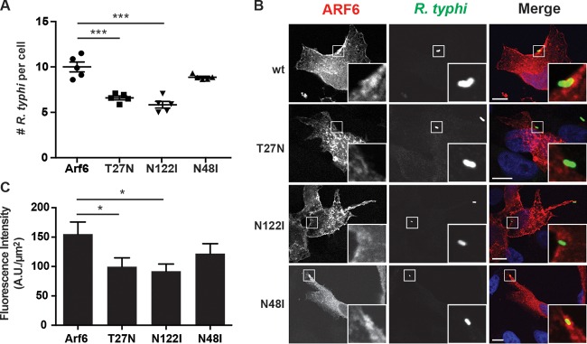
FIG 3.
Inhibition of Arf6 impairs R. typhi infection. (A) Arf6T27N and Arf6N122I decrease R. typhi infection. HeLa cells transiently expressing the mRFP-Arf6 wt or T27N, N122I, or N48I mutant were incubated with partially purified R. typhi (MOI, 100:1) for 2 h at 34°C. The cells were fixed with 4% PFA, and R. typhi was detected with rat anti-R. typhi serum and anti-rat Alexa Fluor 488 antibody. The number of R. typhi bacteria per cell was counted for 100 cells per well. Means ± SEM from five wells of two independent experiments are plotted. ***, P < 0.0001 by one-way ANOVA and Dunnett's multiple-comparison test. (B) Arf6T27N and Arf6N122I do not localize to R. typhi entry foci. HeLa cells were treated as described in panel A, except the incubation time was 15 min. The cells were fixed with 4% PFA, and R. typhi was detected with rat anti-R. typhi serum and anti-rat Alexa Fluor 488 antibody (green). DAPI (blue) is shown in the merged image. Boxed regions are enlarged to show detail. Scale bars, 10 μm. (C) Quantification of Arf6 localization at the R. typhi entry foci. mRFP-tagged Arf6 wt- or T27N, N122I, or N48I mutant-expressing cells were infected with R. typhi and processed as described for panel B. Fluorescence immediately surrounding R. typhi was measured using ImageJ (NIH) and expressed as arbitrary units (A.U.) per square micrometer. The means ± SEM of 10 to 15 bacteria from two independent experiments are plotted. *, P < 0.05 by one-way ANOVA and Dunnett's multiple-comparison test.

