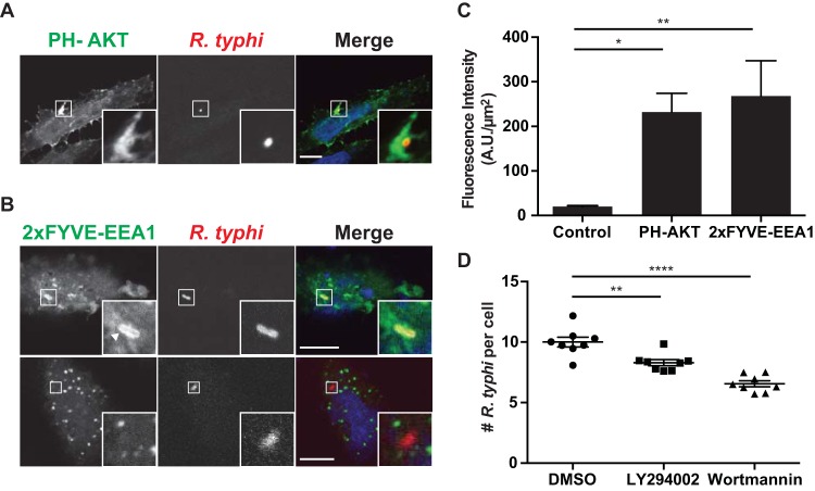
FIG 5.
Recruitment of PI(3,4,5)P3 and PI(3)P to R. typhi entry foci. HeLa cells overexpressing (A) the pleckstrin homology (PH) domain of RAC-alpha serine/threonine-protein kinase (AKT), a PI(3,4,5)P3 and PI(3,4)P2 biosensor (71), or (B) the FYVE domain of early endosome antigen 1 (EEA1), a PI(3)P biosensor (71), were incubated with R. typhi (MOI, 100:1) for 5 min (top panel) or 15 min (bottom panel). Cells were fixed with 4% PFA, and R. typhi was detected with rat anti-R. typhi antibody and anti-rat Alexa Fluor 594 antibody (red). DAPI (blue) is shown in the merged image. Boxed regions are enlarged to show detail. Scale bars, 10 μm. (C) Quantification of PH-AKT and 2× FYVE-EEA1 localization at the R. typhi entry foci. GFP-, PH-AKT-, and 2 × FYVE-EEA1-expressing cells were infected with R. typhi and processed as described for panels A and B. Fluorescence immediately surrounding R. typhi was measured using ImageJ (NIH) and expressed as arbitrary units (A.U.) per μm2. The means ± SEM of 10 to 15 bacteria from two independent experiments are plotted. *, P < 0.05, and **, P < 0.001, by one-way ANOVA and Dunnett's multiple-comparison test. (D) Inhibition of PI3K decreases R. typhi infection. HeLa cells pretreated for 2 h with the PI3K inhibitor LY294002 (5 mM) or wortmannin (100 nM) were infected with R. typhi (MOI, 100:1). Cells were fixed with 4% PFA, and R. typhi was detected with rat anti-R. typhi serum and anti-rat Alexa Fluor 488 antibody and cell membrane stained with wheat germ agglutinin-Alexa Fluor 594 conjugate. The number of R. typhi bacteria per cell for 100 cells in four different wells per condition was calculated for three independent experiments, and means ± SEM are plotted. **, P < 0.01, and ****, P < 0.0001, by one-way ANOVA and Dunnett's multiple-comparison test.

