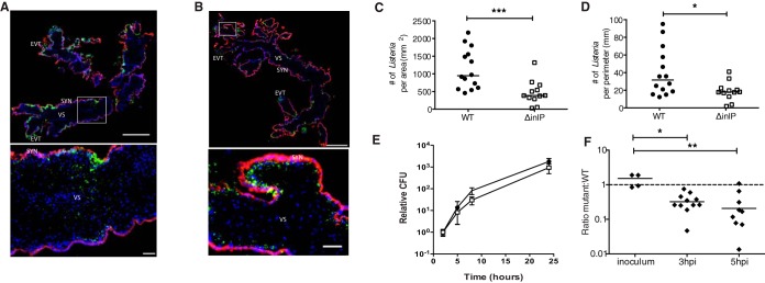
FIG 6.
InlP is important for virulence in human placenta. (A to D) Human placental organ cultures from 3 donors were analyzed 72 h p.i. with either wild type (WT) or ΔinlP mutant. (A) Representative image of a WT-infected placental organ culture stained for L. monocytogenes (green), βHCG (human chorionic gonadotropin) (to stain syncytiotrophoblasts) (red), and DAPI (blue). The rectangle in the top image denotes the location of the high-power image at the bottom. SYN, syncytiotrophoblast; EVT, extravillous trophoblast; VS, villous stroma. (B) Representative image of a placental organ culture infected with ΔinlP mutant and stained as described above for panel A. (C and D) Numbers of L. monocytogenes bacteria present per area (C) and perimeter (D) of villous stroma quantified by immunofluorescence microscopy for 14 WT- and 12 ΔinlP mutant-infected organ cultures. (E) Intracellular growth of the WT (black circles) and ΔinlP mutant (empty squares) in primary human placental fibroblasts as determined by a gentamicin protection assay (averages of data from two independent experiments performed in triplicate). Error bars indicate standard errors of the means. (F) Competitive index in differentiated primary human trophoblast progenitor cells infected with erythromycin-resistant WT and erythromycin-susceptible ΔinlP mutant bacteria at a 1:1 ratio. Mann-Whitney U test was used for statistical analysis of data in panels C to E, and a Kruskal-Wallis test with multiple comparisons was used for analysis of data in panel F (*, P < 0.05; **, P < 0.005; ***, P < 0.001). Horizontal bars indicate medians. The dashed line represents a ratio of 1. Bars, 500 μm (low-power images) and 50 μm (high-power insets).

