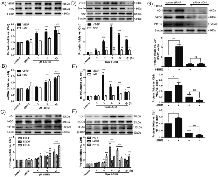
Figure 4. Nrf2 activator t-BHQ activates VEGF via Nrf2/HO-1/HIF-1α pathways.
(A,B) After the BMECs was incubated with 1–10 μM of t-BHQ for 6 h, the mRNA and protein levels of Nrf2 and VEGF was determined using β-actin as the loading control by Western blot and qRT-PCR. The histograms show the ratio of Nrf2/β-actin and VEGF/β-actin. (C) Western blot shows the upregulation of Nrf2 downstream protein HO-1, NQO1 and HIF-1α in response to t-BHQ. (D,E) 10 μM t-BHQ was used to stimulate the BMECs at different time points, and the protein and mRNA levels of VEGF and Nrf2 were measured. (F) The expression of HO-1, NQO1, and HIF-1α were also measured and calculated. (E) After 48 h of transfection with siRNA–HO-1 or control siRNA, the BMECs were incubated with 10 μM t-BHQ for 6 h. Then, Western blot was used to analyse the expression of HO-1, VEGF and HIF-1α. *p < 0.05; **p < 0.05; ***p < 0.001 versus the control siRNA.

