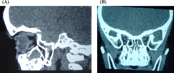
Figure 3.

Patient's craniometaphyseal dysplasia. Cranial computed tomography on diagnosis: Notable abnormalities in bone density and thickness of cranial vault, differentiated abnormalities in bone layers. Bone thickening around nasal pyramid and lamina cribrosa. (A), Sagittal plane; (B), Coronal plane.
