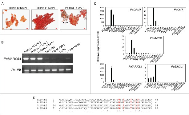
Figure 1.
(A) Acetocarmine staining images showing pollinia at 0, 1, and 3 d after pollination (DAP). Arrowheads point to the pollen tubes. Scale bar = 20 μm. (B) Semi-quantitative RT-PCR showing expression of PaMADS63 mRNA in the indicated tissues. PaUBI1 was used as an internal control. PLB, Protocorm-like-body. (C) Quantitative RT-PCR showing expression patterns of PaDRM1, PaCMT1, PaSUVR1, PaIAA30L1, and PaEIN3L1 in interior tissues of developing ovaries from 30 to 200 d after pollination (DAP). Small-sized PLB (PLBS), medium-sized PLB (PLBM), large-sized PLB (PLBL), 10-day-old protocorms (protocorm10), 20-day-old protocorms (protocorm20), and 30-day-old protocorms (protocorm30). (D) Alignment of the WIYLD domain of Arabidopsis SUVR proteins and PaSUVR1. AtSUVR1, AT1G04050; AtSUVR2, AT5G43990; AtSUVR4, AT3G04380.

