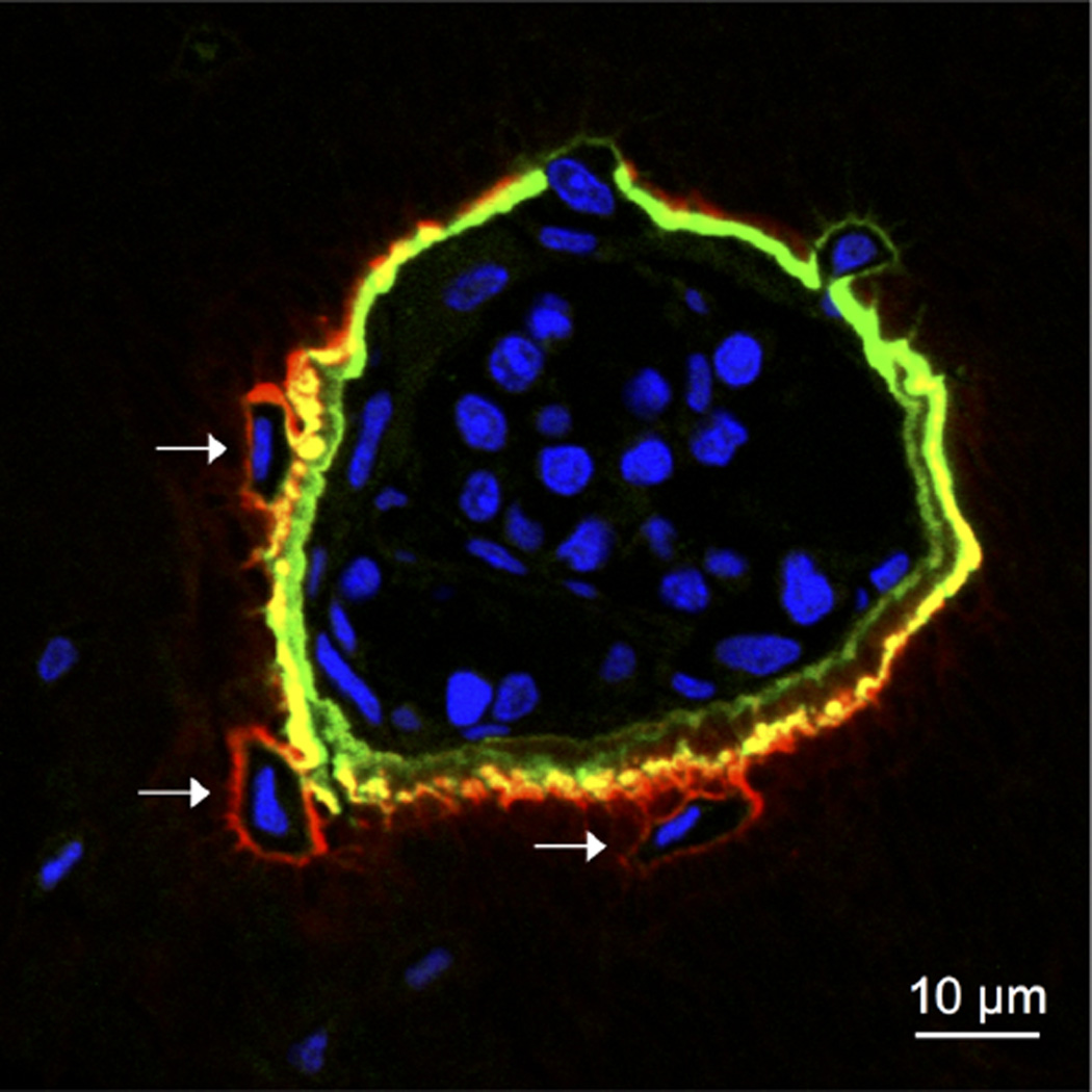
Figure 4.
Localization of a high HAP affinity N-BP drug probe (5(6)-FAM-RIS, green) and a low HAP affinity PC derivative (RhR-RISPC, red) in the bone of a 9-week-old male Sprague–Dawley rat (0.2 kg). 5(6)-FAM-RIS (0.345 mg/kg) and 5(6)-RhR-RISPC (0.385 mg/kg) were administered subcutaneously via single dose, and 7 days later their localization pattern was analyzed in transverse histological sections of the tibia. Nuclei were stained with DAPI (blue). Shown is a pocket of bone marrow surrounded by labeled bone matrix, with several newly formed osteocytes indicated by arrows. Note the differential labeling pattern of the two compounds with the lower affinity compound (5(6)-RhR-RISPC) penetrating more deeply into the bone matrix and appearing more abundantly in the osteocyte lacunae (arrows).

