Fig. 4.
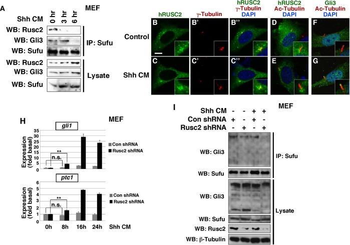
Rusc2 inhibits signaling-induced dissociation of Sufu-Gli protein complexes. (A) CoIP showing sequential dissociation of Rusc2-Sufu-Gli3 complexes in MEFs upon Shh-conditioned medium treatment. Endogenous Sufu was immunoprecipitated. The amount of endogenous Gli3 and Rusc2 associated with Sufu was assessed by western blot. (B-G) Confocal images showing the subcellular localization of FLAG-hRUSC2 in control (upper row) and Shh-conditioned medium-stimulated (bottom row) cells. (B,C) Anti-FLAG staining for hRUSC2. (B′,C′) γ-tubulin staining. (B″,C″) Merges of B,B′ and of C,C′. (D,E) Merged images of hRUSC2 and acetylated-tubulin staining. (F,G) Merged images of Gli3 and acetylated-tubulin staining. Insets are higher magnification views of the area around cilia. Scale bar: 10 µm. (H) RT-PCR showing the expression of Gli1 and Ptc1 in unstimulated MEFs and MEFs treated with Shh-N-conditioned medium for 8, 16 and 24 h. Data are shown as mean±s.d. **P<0.01. n.s., non-significant. (I) CoIP experiments to assess the effect of Rusc2 knockdown on Shh-induced dissociation of Sufu-Gli3 protein complexes in MEFs. Endogenous Sufu and Gli3 were analyzed. The dose of Shh-N-conditioned medium used was identical to that in H.

