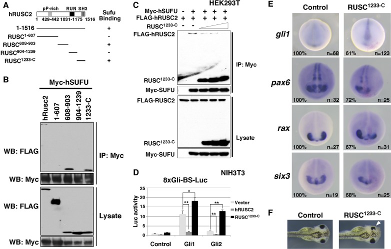
Fig. 7.
Dominant-negative Rusc enhances Hh signaling in Xenopus embryos and impairs eye development. (A) Schematic of hRUSC2 and deletion derivatives. Whether an hRUSC2 construct interacts with hSUFU in the CoIP experiment is indicated by + or −. (B) CoIP results showing that hSUFU interacts with full-length hRUSC2, RUSC608-903 and RUSC1233-C. (C) CoIP showing that overexpression of RUSC1233-C reduces the binding between hSUFU and full-length hRUSC2. (D) Dual-luciferase assay showing that the activities of Gli1 and Gli2 are enhanced by co-overexpression of RUSC1233-C in NIH3T3 cells. Data are shown as mean±s.d. *P<0.05, **P<0.01. (E) In situ hybridization showing the expression of gli1, pax6, rax and six3 in control (left) and RUSC1233-C overexpression (right) Xenopus embryos at stage 20. At the 8-cell stage, one of the dorsal animal blastomeres was injected with a mixture of RUSC1233-C (1 ng) and n-β-gal (250 pg) encoding RNAs. (F) Overexpression of RUSC1233-C (1 ng) reduced the size of the eye (arrowhead).

