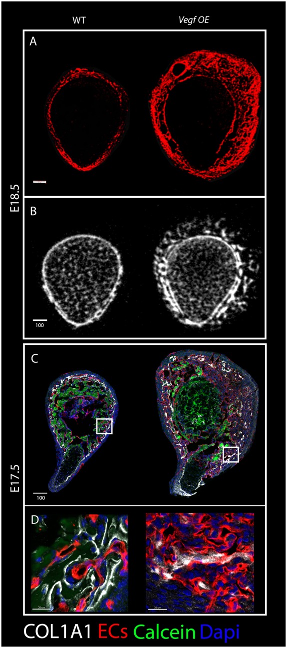
Fig. 10.

Bone malformations upon Vegf overexpression. (A) Cryostat cross-sections of humeri from wild-type and Vegf-OE mice at E18.5 immunostained for blood vessels (red) show expansion of the vascular patterning domain in the mutant. (B) Cross-sectional views of three-dimensional reconstructions from micro-CT scans of humeri from E18.5 wild-type (left) and Vegf-OE (right) mice illustrate the abnormal arrangement of the primordial cortex in the mutant. (C) Cross-sections of E17.5 humeri immunostained for blood vessels (red) and COL1A1 (white) show discontinuous collagen type I distribution in the mutant. (D) Magnifications of the boxed areas demonstrate collagen I association with ECs in both wild type and mutant. Scale bars: 100 µm in A-C; 20 µm in D.
