Fig. 3.
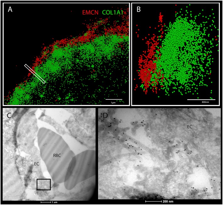
Super-resolution microscopy and transmission electron microscopy show collagen I on ECs in developing bones. (A,B) STORM images of cross-sections of humeri at E15.5 immunostained for EMCN (red) and collagen I (green). (B) A side view of the boxed area in A. (C,D) Cross-sections of E16.5 mouse humerus observed by TEM. (C) Cross-sections of blood vessels from the bone area. (D) Magnification of the boxed area in C. Collagen type I immunostained with 10 nm gold particles is located on the endothelial cell (EC) surface. RBC, red blood cells. Scale bars: 1 µm in A; 800 nm in B; 1 µm in C; 200 nm in D.

