Fig. 8.
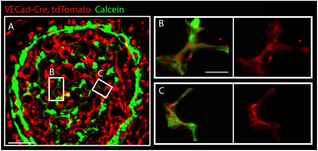
Low expression of an EC marker in newly mineralized areas. (A) Cross-section of humerus from VECad-Cre, tdTomato (red) mouse embryo at E17.5 injected with calcein (green). (B,C) Magnifications of the boxed areas in A show low expression of VECad by ECs in areas of newly deposited mineral, indicated by weak calcein signal. Scale bars: 100 µm in A; 30 µm in B,C.

