Fig. 1.
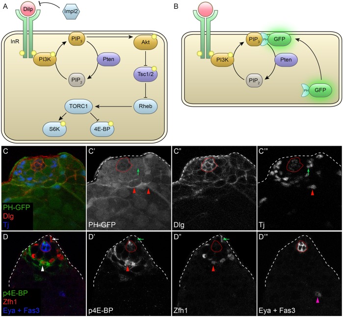
PI3K and Tor activity are observed during CySC differentiation. (A) Simplified model of the PI3K/Tor pathway. Here, PI3K is activated by the insulin receptor (InR) tyrosine kinase following binding of a Drosophila insulin-like peptide (Dilp). InR phosphorylates PI3K, which in turn phosphorylates PIP2 lipids to create PIP3. Pten is a lipid phosphatase that catalyzes the reverse reaction. Subsequently, PIP3 activates Akt, which inactivates the Tor inhibitor Tsc1/2. The TORC1 complex is then activated and leads to the phosphorylation of S6K and 4E-BP. The secreted Dilp antagonist ImpL2 blocks Dilps from binding to InR. Phosphorylation events are indicated by the yellow symbols. (B) The PH-GFP reporter of PIP3 consists of GFP fused to the pleckstrin homology (PH) domain of Grp1, which binds PIP3. This GFP fusion is cytoplasmic, but when PIP3 levels are high it translocates to the plasma membrane. (C) The PI3K reporter PH-GFP (green) was predominantly cytoplasmic in CySCs around the hub, indicating low PI3K activity. Arrow in C′,C‴ marks a CySC with low membrane PH-GFP. Membrane-associated PH-GFP was observed as cyst cells begin to differentiate, one further cell diameter away from the hub. Arrowheads in C′,C‴ mark differentiating cyst cells that have upregulated membrane PH-GFP. Tj (blue and C‴) labels CySCs and early cyst cells, Dlg (red and C″) marks somatic cell membranes. (D) The Tor activity reporter p4E-BP (green) was detected at high levels in somatic cells adjacent to stem cells. Arrow in D-D″ marks a Zfh1-positive CySC next to the niche that has low p4E-BP. Arrowhead in D-D″ labels a Zfh1-positive early CySC daughter cell primed for differentiation that is one cell diameter away from the CySCs and that has high levels of p4E-BP. Arrowhead in D‴ marks an Eya-positive differentiating cyst cell. Fas3 (blue and D‴) marks the hub; CySCs are the Zfh1-positive cells (red and D″) that are closest to the hub. Dotted red line outlines the hub.

