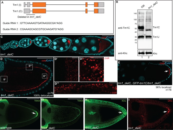
Fig. 6.
Generation of a Tm1C-null mutant. (A) Schematic showing the region deleted in Tm1C-null mutants. We refer to this strain as tm1_delC. Gray boxes represent untranslated regions and orange boxes indicate coding regions. The sequences of the guide RNAs used to generate this deletion are also indicated. (B) Ovarian lysates were prepared from wild-type (lane 1) or the tm1_delC mutant (lane 2). The lysates were analyzed by blotting using the anti-Tm1C antibody. The blot was subsequently stripped and probed with an anti-Khc antibody (bottom panel). (C,D) Ovaries from tm1_delC mutants were processed for smFISH using probes against osk (red) and were counterstained with DAPI (cyan). D′,D″ and D‴ are enlarged images of boxes in D. (E) Ovaries from tm1_delC mutants expressing the GFP–Tm1C transgene were processed for smFISH using probes against osk (red) and were counterstained with DAPI (cyan). The arrow indicates localized osk mRNA. (F,G) Ovaries from wild-type (F) or tm1_delC mutants (G) were fixed and processed for immunofluorescence using an antibody against Vasa. The arrow indicates localization of Vasa to the pole plasm in wild-type egg chambers. (H,I) Ovaries from tm1_delC mutants were processed for immunofluorescence using antibodies against Khc (H) or Dhc (I). Arrows indicate posterior-localized Khc and Dhc in Tm1C-null mutants. Scale bars: 50 μm (C–I); 5 μm (D′–D‴).

