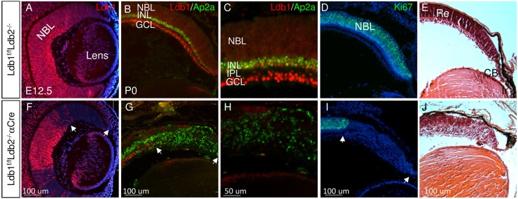
Fig. 1.
Ldb1 and Ldb2 are required for maintaining proliferation and multipotency of RPCs. Gene expression and tissue morphology were monitored in control (A-E) and in Ldb1loxP/loxP;Ldb2−/−;α-Cre (F-J) eyes, determined by immunofluorescence analyses (A-D,F-I) and Hematoxylin and Eosin staining (E,J) for Ldb1 (red in A-C,F-H), Ap2a (green in B,C,G,H) and Ki67 (green in D,I) at E12.5 (A,F) and P0 (B-E,G-J). Counterstaining was with DAPI (blue, A,D,F,I). C,H are higher magnifications of staining for Ldb1 and Ap2a. White arrows in F,G,I mark the mutation area. CB, ciliary body; GCL, ganglion cell layer; INL, inner nuclear layer; IPL, inner plexiform layer; Ir, iris; NBL, neuroblastic layer; Re, retina. Scale bars: 100 μm in A,B,D-G,I,J; 50 μm in C,H.

