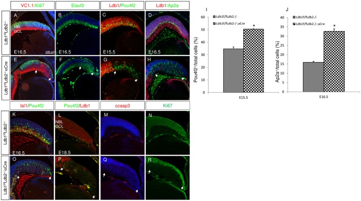
Fig. 3.
Ldb proteins are required first for preventing premature differentiation into ganglion and amacrine lineages and later for ganglion cell survival. (A-H) In control (A-D) and Ldb1loxP/loxP;Ldb2−/−;α-Cre (E-H) retinas, immunofluorescence analyses show the expression of Vc1.1 and Ki67 (red and green; A,E), Elavl3 (B,F), Ldb1 and Pou4f2 (red and green; C,G), and Ldb1 and Ap2a (red and green; D,H). Arrows indicate mutated area. (I) The percentage of Pou4f2+ cells at E15.5 in the control retina (34.7±1.55) and in Ldb1/Ldb2 mutant retina (50.4±0.21). Data are mean±s.d., n=3, *P<0.002 calculated using a two-tailed t-test. (J) The percentage of Ap2a+ cells at E16.5 in the control retina (15.8±0.56%) and in Ldb1/Ldb2 mutant retina (32.6±1.3%). Data are mean±s.d., n=3, *P<0.001 calculated using a two-tailed t-test. (K-R) In control (K-N) and Ldb1loxP/loxP;Ldb2−/−;α-Cre (O-R) retinas, immunofluorescence analyses show the expression of Isl1 and Pou4f2 at E16.5 (red and green, K,O), Pou4f2 and Ldb1 (green and red, L,P), cCasp3 (red, M,Q) and Ki67 (green, N,R) at E18.5. GCL, ganglion cell layer; NBL, neuroblastic layer. Scale bar: 100 μm.

