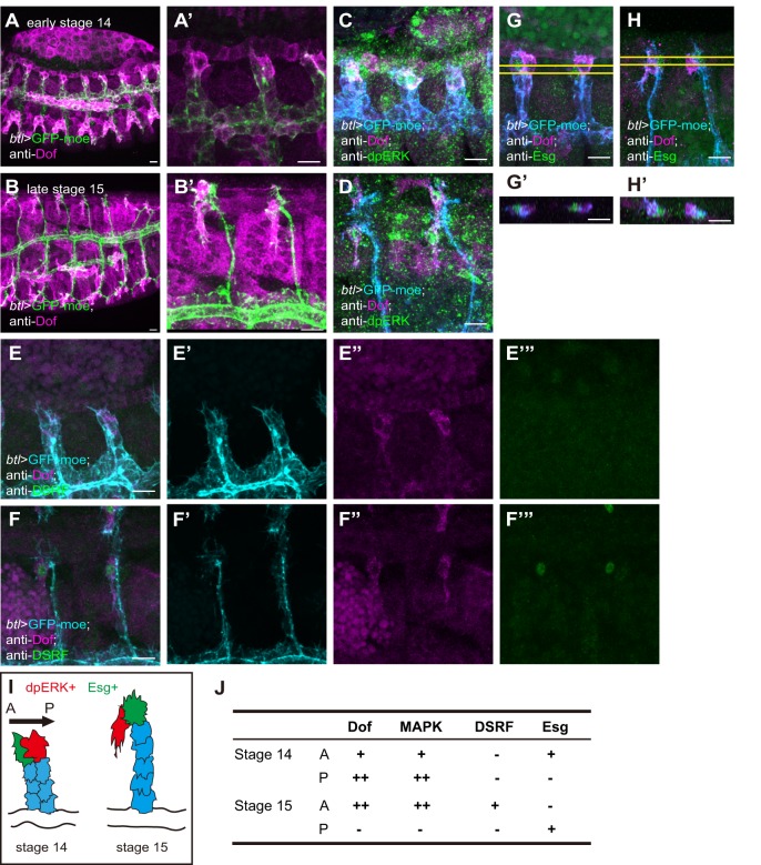
Fig. 1.
Normal dorsal branch (DB) development in Drosophila embryos. Anti-Dof staining (A-B′) and co-staining for Dof and dpERK (C,D), DSRF (E-F‴) or Esg (G-H′) of btl-Gal4-driven UAS-GFP-moe Drosophila embryos. (A,C,E,G) Early stage 14. (B,D,F,H) Late stage 15. (A′,B′) Magnified views of A,B. (E′-E‴,F′-F‴) Single-channel images of E,F. (G′,H′) x-z views of the area between the yellow lines in G,H. Scale bars: 10 µm. (I) Schematic of the DB at stages 14 and 15, showing Esg-positive cells (green), dpERK-positive cells (red) and stalk cells (SCs; blue). (J) Summary of gene expression patterns in DB tip cells. A, anterior; P, posterior.

