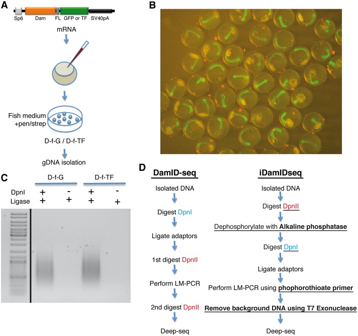
Fig. 2.
Modification of the crucial steps of the DamID protocol. (A) Medaka zygotes were injected with mRNA coding for Dam-f-GFP or Dam-f-TF (Medaka Rx2). Embryos were maintained in ERM supplemented with an antibiotic solution and gDNA was isolated at stage 22. (B) Medaka embryos (stage 22) expressing Dam-f-GFP. (C) DamID LM-PCR at 25 cycles using the modifications presented in the main text generates only DpnI-dependent amplification (see Materials and Methods, iDamIDseq protocol). (D) Flowchart comparing the standard DamID-seq protocol (based on Wu et al., 2016) with the iDamIDseq protocol (improvements are underlined).

