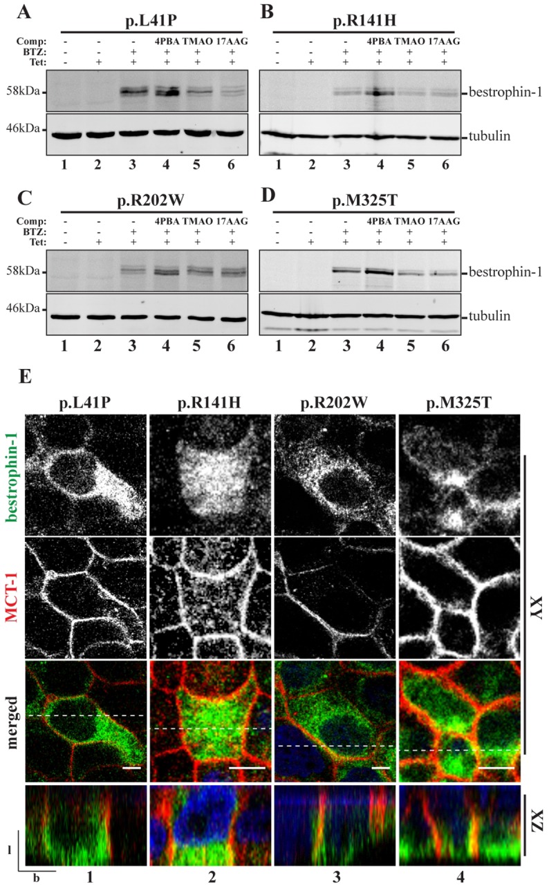
Fig. 2.

Small molecule treatment of ARB-associated bestrophin-1 in stable polarised MDCKII cells. (A-D) Mutant bestrophin-1 proteins expression level was investigated by western blot. p.L41P, p.R141H, p.R202W and p.M325T bestrophin-1 expression was induced with tetracycline (Tet) and cells were treated with BTZ or a combination of BTZ+4PBA, TMAO or 17-AAG for 24 h before direct lysis in SDS sample buffer. An anti-tubulin antibody was used as a loading control. (E) Confocal immunofluorescence analysis was used to investigate mutant bestrophin-1 localisation (green) in MDCKII stable cells lines following 24 h treatment with BTZ+4PBA. Representative XY and YZ scans for each mutant are shown. Co-localisation with MCT-1 (red) was used as a marker for correct trafficking to the basolateral plasma membrane. Scale bar: 5 µm. l, lateral; b, basal. Dotted line in merged images shows position of the XZ scan.
