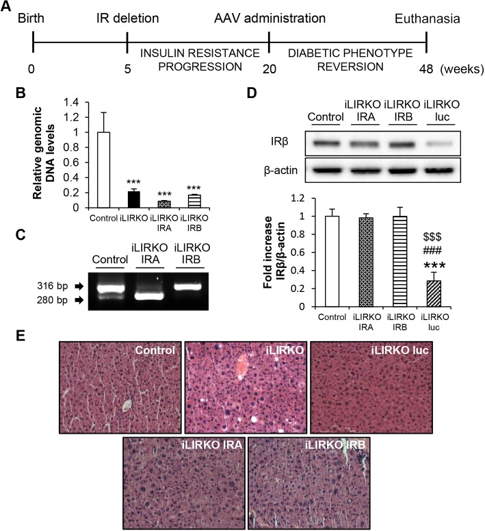
Fig. 4.
AAV-mediated IRA and IRB expression in the liver recovered initial levels of insulin receptor. (A) Schedule of AAV administration experiments. (B) Mouse genomic DNA Insr exon 4 levels were analysed by qPCR in 9-month-old control and iLIRKO mice with or without AAV-IRA or AAV-IRB administration. (C) Mouse Insr isoforms in control and human INSR isoforms in iLIRKO IRA and iLIRKO IRB were analysed by RT-PCR in livers from 9-month-old mice. (D) Representative IRβ expression analysed by western blot in liver homogenates from 9-month-old control, iLIRKO IRA, iLIRKO IRB and iLIRKO luc mice. β-actin was used as loading control. The histogram shows the band intensity quantification. Data are means±s.e.m. for each experimental group, n=5. (E) Representative H&E staining of liver sections from 9-month-old control, iLIRKO, iLIRKO luc (upper panels) and iLIRKO IRA, iLIRKO IRB (lower panels) mice. Image magnification: 20×. ***P<0.001 versus control mice; ###P<0.001 versus iLIRKO IRA; $$$P<0.001 versus iLIRKO IRB by one-way ANOVA with Bonferroni post test.

