Fig. 1.
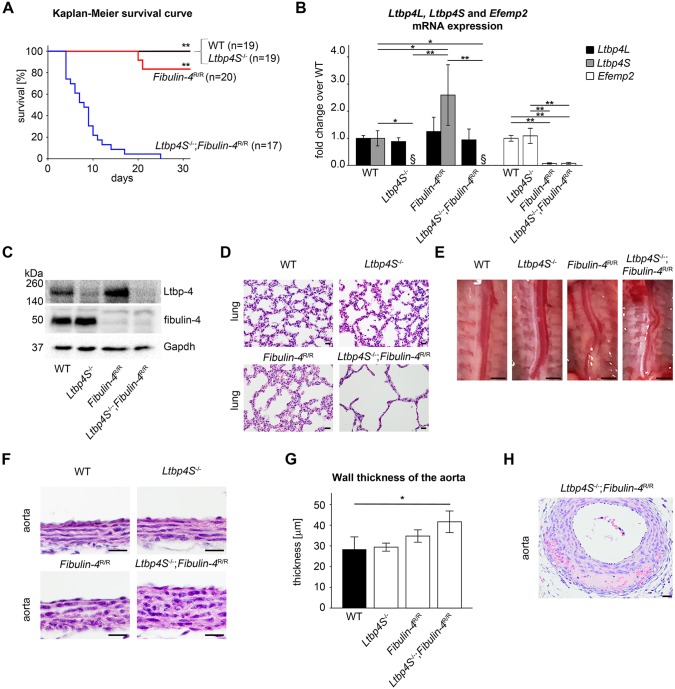
Clinical and pathological findings of Ltbp4S−/−;Fibulin-4R/R mice with reduced interaction of Ltbp-4L and fibulin-4. (A) Kaplan–Meier survival curve revealed significantly higher neonatal mortality in Ltbp4S−/−;Fibulin-4R/R mice (n=17) compared to WT (n=19), Ltbp4S−/− (n=19) and Fibulin-4R/R (n=20) mice, which showed no differences in survival. Differences between groups were analyzed by log-rank test (**P<0.01 vs Ltbp4S−/−;Fibulin-4R/R). (B) Quantitative PCRs and (C) representative immunoblots showed Ltbp-4 and Efemp2 mRNA and Ltbp-4 and fibulin-4 protein expression of WT (n=3), Ltbp4S−/− (n=3), Fibulin-4R/R (n=4) and Ltbp4S−/−;Fibulin-4R/R (n=4) lungs. Differences between groups were analyzed by two-way ANOVA, followed by Bonferroni correction (*P<0.05 and **P<0.01; §below detection limit). (D) In Ltbp4S−/− and Fibulin-4R/R mice, the pulmonary parenchyma had enlarged alveolar spaces with reduced numbers of alveoli compared to WT mice. Lungs of Ltbp4S−/−;Fibulin-4R/R mice showed a higher degree of severely enlarged alveolar spaces with multifocal areas of atelectasis and a lack of alveolar and lobular architecture compared to all other genotypes. Scale bars: 20 µm. (E) Ltbp4S−/−, Fibulin-4R/R and Ltbp4S−/−;Fibulin-4R/R mice displayed tortuous aortas. Scale bars: 10 mm. (F,G) Abdominal aortas showed marked thickening of the aortic wall in Ltbp4S−/−;Fibulin-4R/R mice compared to the other genotypes. Differences between groups were analyzed by two-way ANOVA, followed by Bonferroni correction (n=3; *P<0.05; scale bars: 20 µm). (H) Abdominal aortas of Ltbp4S−/−;Fibulin-4R/R mice showed intramural hemorrhages and destruction of the aortic wall with necrotic cellular debris (scale bar: 20 µm). Data are presented as means±s.d.; n indicates the number of analyzed tissue of individual mice or analyzed mice.

