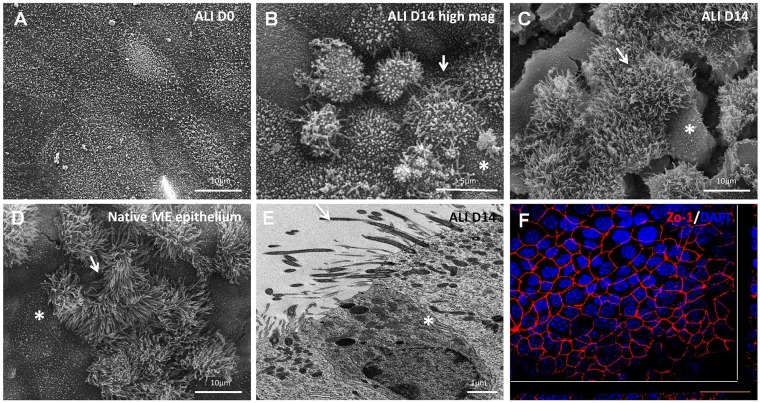
Fig. 2.
Electron microscopy of mMEC cultures. (A-D) Scanning electron microscopy of ALI day 0 mMEC cultures showing large flat polygonal cells with apical microvilli (A), ALI day 14 cultures showing dome shaped cells at higher magnification (B) and combination of interspersed flat polygonal and densely ciliated cell populations a lower magnification (C) resembling the morphology of native middle ear epithelium (D). Cracks in the membrane are due to processing of samples for s.e.m. White arrows mark elevated ciliated cells and asterisks mark flatter polygonal cells. (E) Transmission electron microscopy of ALI day 14 mMEC cultures showing adjacent ciliated and secretory cells and formation of tight junctions demonstrated by presence of desmosomes (asterisk). Arrow shows cilia. (F) Immunofluorescence confocal microscopy image showing formation of tight junctions marked by ZO-1-positive staining. Cross-sections through both axes of the membrane are shown beneath and to the right beyond the white lines. Images are representative of three independent batches. Scale bars: 10 μm in A,C,D; 5 μm in B; 1 μm in E; 50 μm in F.

