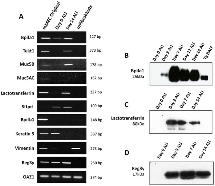
Fig. 3.
Expression of epithelial markers in mMEC cultures. (A) End-point RT-PCR showing expression of a selected panel of upper airway-associated genes in mMEC original cells isolated from the middle ear cavity, ALI day 0 cells and ALI day 14 cells, with fibroblasts as a negative control. Expression profile of ALI day 14 cells was similar to mMEC original cells isolated from the middle ear for most genes. (B-D) Detection of Bpifa1 (B), lactotransferrin (C) and Reg3γ (D) in the apical washes from differentiating cells using western blotting. Data is representative of three independent cultures.

