Fig. 4.
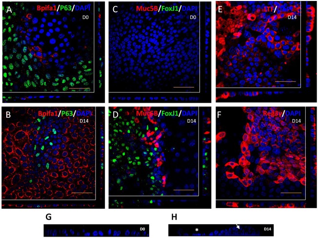
Localisation of epithelial markers in mMEC cultures. (A,C) Immunofluorescence confocal images (representative of three independent batches) showing abundant expression of the basal cell marker, P63; limited expression of the secretory protein, Bpifa1 (A), and no expression of goblet cell marker, Muc5B and the ciliated marker, FoxJ1 (C) in undifferentiated ALI day 0 mMEC cultures. (B,D,E,F) Differentiated mMEC ALI day 14 cultures showing expression of secretory cells positive for Bpifa1 (B), lactotransferrin (E), Reg3γ (F), goblet cells positive for Muc5B and ciliated cells positive for FoxJ1 (D). Cross-sections through both axes of the membrane are shown beneath and to the right beyond the white lines. (G,H) High magnification z-stack cross sections of nuclei stained with DAPI shows that ALI day 0 cells form a flat monolayer (G) whereas ALI day 14 cells are a combination of pseudostratified elevated cells (arrow) and flatter cells (asterisk) (H). Scale bars: 50 μm.

