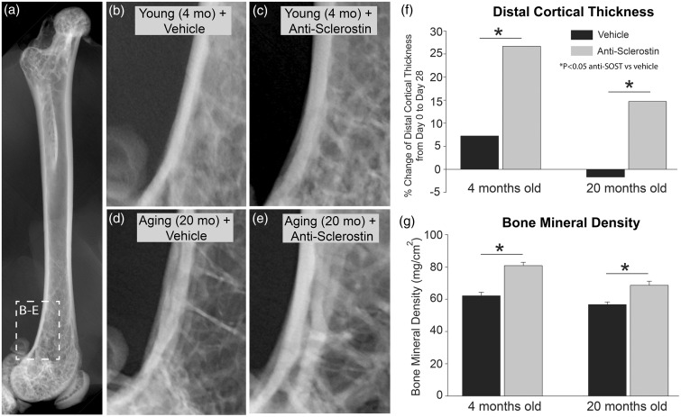
Figure 1.
Anti-sclerostin increases the thickness of cortical bone and bone mineral density in young and geriatric mice. (a) Anterior/posterior (AP) radiograph of a naïve young (4 months old) male C57Bl/6 mouse shown for orientation. (b–d) Representative radiographs of the distal end of the femur of young (4 months old) (b, c) and aging (20 months old) (d, e) mice showing bone following of vehicle (b, d) or anti-sclerostin (25 mg/kg, i.p.) (c, e) for 28 days. (f) Histogram showing the percent change of thickness of the cortical bone in the distal head of the femur after 28 days of vehicle vs. anti-sclerostin treatment. Note that following 28 days administration of vehicle alone, the cortical thickness increased in young adult animals and decreased slightly in aging animals. In contrast, in both young and geriatric animals administered anti-sclerostin, there was a significant increase in the thickness of cortical bone. (g) Anti-sclerostin therapy also increased bone mineral density (BMD) in the distal head of the femur compared to administration of the vehicle alone. Bars represent mean ± SEM; p < 0.05 after a one-way ANOVA, Tukey’s post-hoc test.

