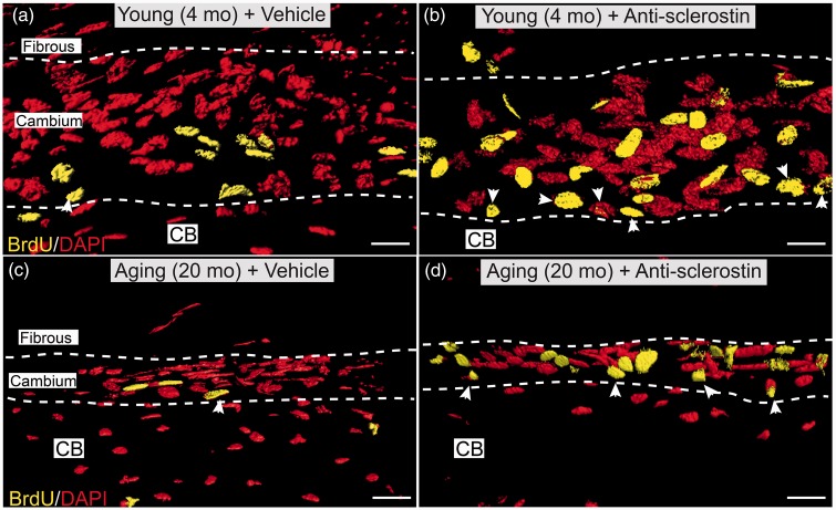
Figure 3.
Confocal images showing that administration of anti-sclerostin increases the number of BrdU+ cells in the cellular “cambium” layer of the periosteum. (a–d) Representative high-power confocal images (3D-rendering performed with Imaris software) of decalcified frozen section of the cambium layer from distal mid-diaphysis of the femur from the young (4 months old) (a, b) and aging (20 months old) (c, d) animals administered vehicle (PBS) (a, c) or anti-sclerostin (25 mg/kg, i.p.) (b, d) every five days for 28 consecutive days. During the last seven days (days 21–28) animals were implanted with a mini-osmotic pump which provided continuous systemic release of BrdU. After decalcification and cutting frozen section of bone, sections stained with an antibody raised against BrdU (yellow; marker for cellular proliferation) and counterstained with DAPI (red; marker for nuclei). Note the increased number of BrdU+ cells in the cambium layer of anti-sclerostin (b, d) vs. vehicle (a, c) treated animals. Also note the increased number of BrdU+ cells at the interface between the cambium layer and cortical bone was termed the bone-lining layer (arrow heads). Scale bar, 30 µm. CB: cortical bone.

