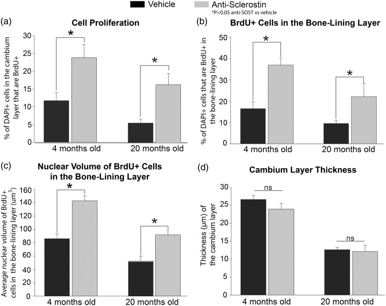
Figure 5.
Histograms showing that administration of anti-sclerostin increases the proliferation of cells in the cambium layer of the periosteum. (a–d) Histograms illustrating the changes to the periosteum with administration of anti-sclerostin (25 mg/kg, i.p.; gray bars) or vehicle (PBS; black bars) every five days for 28 consecutive days to young (4 months old) and aging (20 months old) animals. (a) There is a significant increase in cell proliferation (percentage of DAPI cells marked with BrdU) in the cambium layer after administration of anti-sclerostin in both young and old animals compared to vehicle treated animals. (b) Anti-sclerostin-treated animals had an increase in the total number of DAPI+ cells that were BrdU+ cells at the bone-lining zone in both young and old animals compared to vehicle-treated animals. (c) Administration of anti-sclerostin increased nuclear volume of BrdU+ at the bone-lining surface in young and old animals compared to vehicle-treated animals. (D) Administration of anti-sclerostin has no effect on the cambium layer thickness compared to vehicle-treated animals in either age groups. Bars represent mean ± SEM; p < 0.05 after a one-way ANOVA, Tukey’s post-hoc test.

