Figure 2.
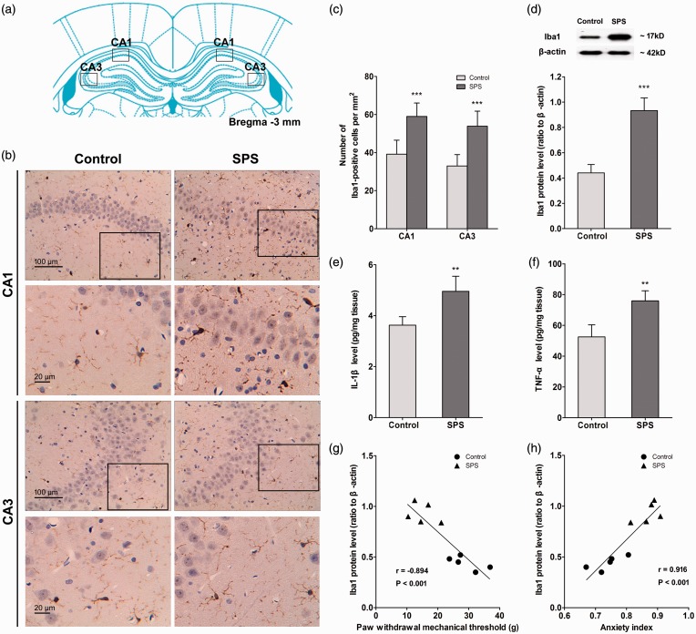
Single prolonged stress (SPS) induced activation of microglia and accumulation of pro-inflammatory cytokines in the hippocampus. After the last behavioral testing on day 7 after SPS, the brain samples of rats were collected. Hippocampal microglia were visualized using Immunohistochemistry, and images were captured under a light microscope. (a) The location of image capture.34 (b) The images were captured in the CA1 and CA3 regions of the hippocampus. (c) The number of Iba1-positive cell bodies was counted using ImageJ software (n = 10 sections of three rats for each group). (d) Iba1 protein levels in the hippocampus were measured using Western blot analysis, and hippocampal levels of IL-1β (e) and TNF-α (f) were measured using enzyme-linked immuno sorbent assay analysis (n = 5 in each group). Correlation analysis between hippocampal Iba1 expression level and PWMT (g) as well as anxiety index (h) on day 7 was conducted (n = 5 in each group). Data were expressed as the mean ± standard deviation. **P < 0.01, ***P < 0.001 compared with control group.

