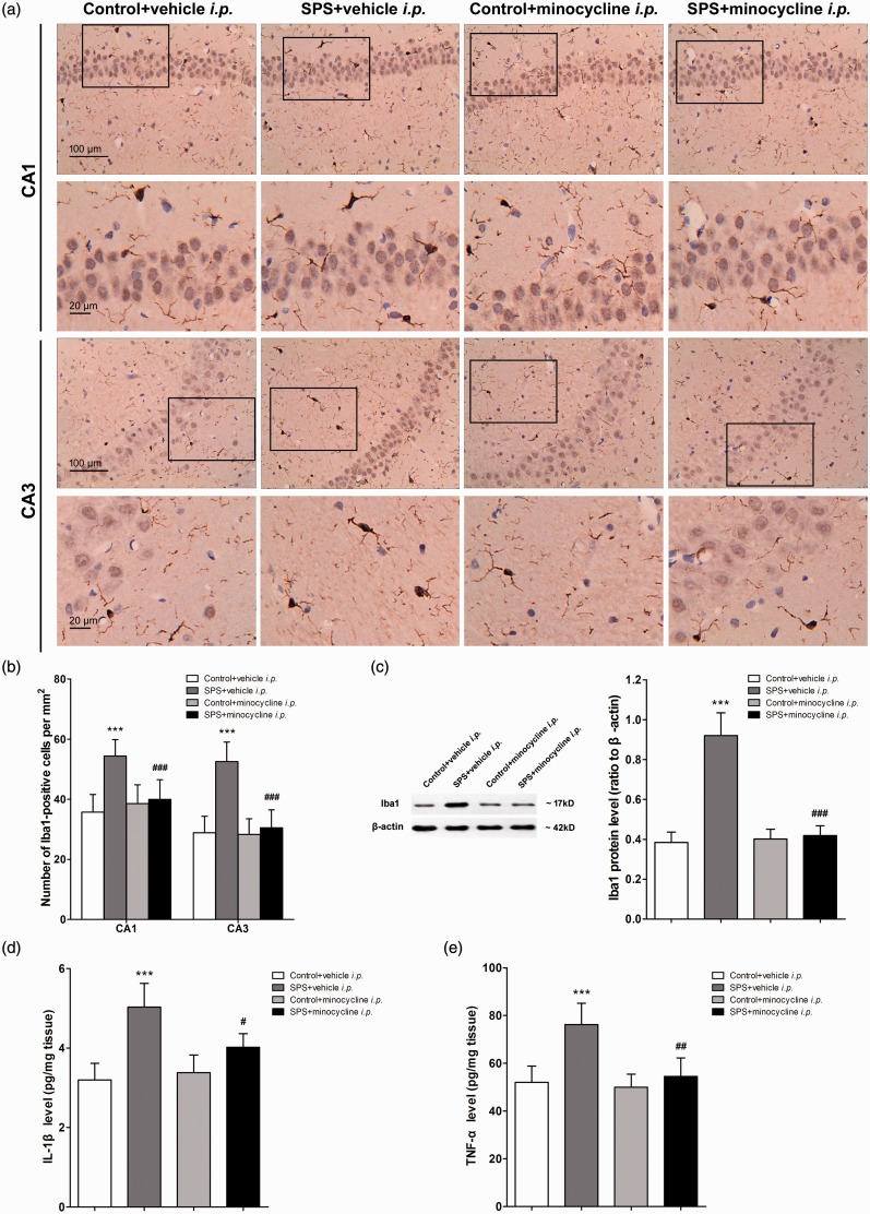
Figure 4.
Intraperitoneal (i.p.) injection of minocycline suppressed hippocampal microglia activation and inflammatory cytokine accumulation in rats exposed to single prolonged stress (SPS). After the last behavioral testing on day 7 after SPS, the brain samples of rats were collected. Hippocampal microglia were visualized using immunohistochemistry. (a) The images were captured in the CA1 and CA3 regions of the hippocampus. (b) The number of Iba1-positive cell bodies was counted using ImageJ software (n = 10 sections of three rats for each group). (c) Iba1 protein levels in the hippocampus were measured using Western blot analysis, and hippocampal levels of IL-1β (d) and TNF-α (e) were measured using enzyme-linked immuno sorbent assay analysis (n = 5 in each group). Data were expressed as the mean ± standard deviation. ***P < 0.001 compared with group control + vehicle i.p.; #P < 0.05, ##P < 0.01, ###P < 0.001 compared with group SPS + vehicle i.p.

