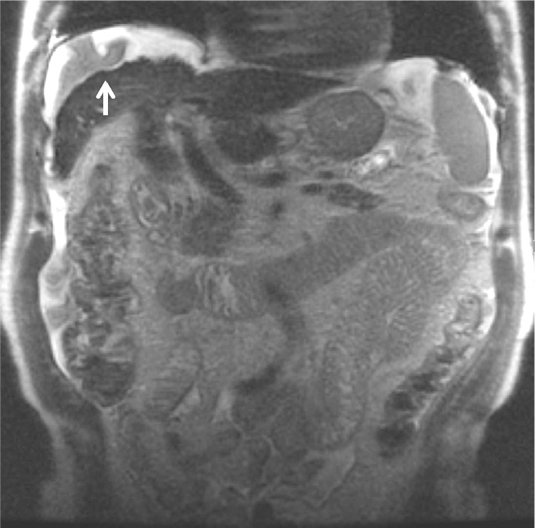
Figure 2.
SSFSE T2-weighted, coronal (TE = 99 milliseconds, slice thickness = 4 mm). Coronal T2-weighted images are useful to obtain an overview of the upper abdomen, in particular, to evaluate the extension of fluid collections. In this 84-year-old man with liver cirrhosis, a perihepatic collection is present, with artifacts due to fluid movements (white arrow).

