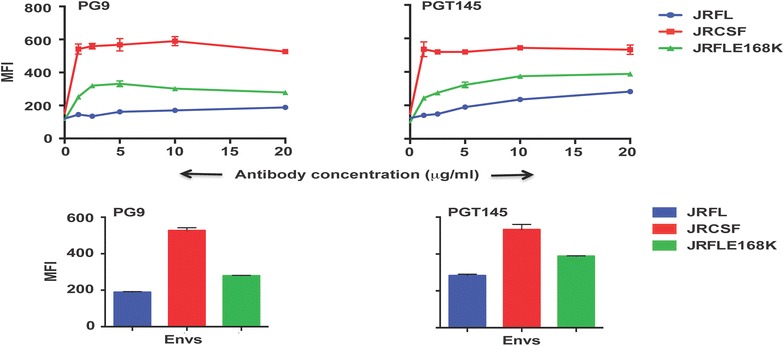
Fig. 3.

Comparative binding of JRFL, JRCSF and JRFL (E168K) to V2 “cap” targeted glycan and conformation-dependent Abs PG9 and PGT145 over a range of antibody concentrations by FACS-based cell surface staining assay. The bar diagram represents the binding of the respective antibodies to wild type and mutant Envs at 20 µg/ml of antibody concentration
